

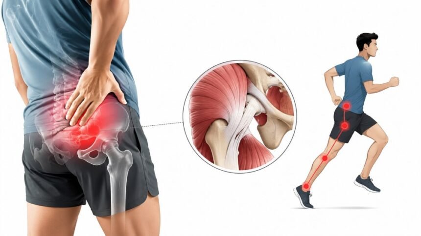
Try this before you read any further. Without thinking about it, put your hand on your hip.
Almost everyone lands in the same place, on the hard bony ridge at the side of the leg, roughly level with a trouser pocket. That ridge is the greater trochanter. It is a real structure, and it causes real pain, but it is not the hip joint. The joint sits deeper and further forward, tucked behind a considerable amount of muscle, and when the joint itself is the problem, the pain very often shows up somewhere your hand never went.
That single mismatch, between where a structure lives and where it hurts, explains an enormous amount of the confusion around hip complaints. It also explains why people spend months faithfully treating the wrong thing, sometimes with surgery.
Joint Is Deeper Than The Pain
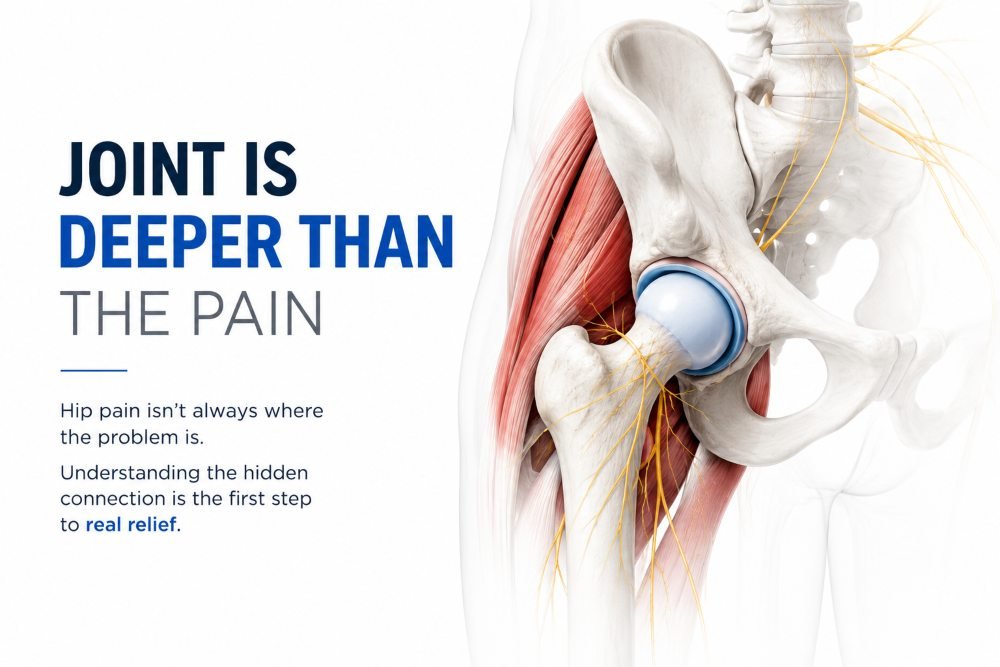
The hip is a ball-and-socket joint, formed by the head of the femur sitting inside the acetabulum of the pelvis, wrapped in a capsule, rimmed by a ring of cartilage called the labrum, and buried under a thick envelope of muscle. Almost none of it can be touched from the outside.
Just as importantly, the nerve supply to the joint does not map neatly onto the skin above it. The capsule receives branches from several nerves, and those nerves also serve territory well away from the joint. The brain, receiving a signal it cannot precisely localise, assigns it to somewhere plausible. Often that somewhere is wrong.
MedlinePlus, the National Library of Medicine’s patient-facing resource, makes the point plainly in its overview of hip pain: pain felt around the hip may actually be coming from the back, and pain from the joint itself is most often felt in the groin.
What The Research Actually Shows About Location
This is not folklore. It has been measured, and the measurements are more surprising than most patients expect.
The cleanest study of the question took fifty-one patients with a hip joint confirmed as the pain source through fluoroscopically guided intra-articular injection. It mapped where each of them felt their pain beforehand. Groin involvement appeared in 55 percent and thigh involvement in 57 percent, which is roughly what the textbooks predict. The full pain-mapping results also found something that contradicts the textbooks: buttock pain appeared in 71 percent of patients, making the buttock the single most common referral area from a symptomatic hip joint, and 22 percent of patients had pain extending below the knee.
Read that again if you have ever been told that buttock pain means a back problem. It very often does. But it is also the most frequent place a bad hip announces itself.
A separate British study looked specifically at pain radiating below the knee, comparing patients waiting for hip replacement against patients waiting for spinal decompression. The finding worth carrying into a consultation is that below-knee pain from hip disease is real and reasonably common, despite a widespread clinical assumption that it does not happen.
Both studies point the same direction. The location of hip pain is a clue, not a verdict.
Reading The Map
With that caveat firmly in place, the patterns are still worth knowing, because they shape the questions a clinician will ask.
- Groin pain points inward, toward the joint. MedlinePlus lists arthritis as typically felt in the front of the thigh or the groin, and describes femoroacetabular impingement as abnormal bone growth around the hip that precedes arthritis and produces pain with movement and exercise. The American Academy of Orthopaedic Surgeons goes further in its explainer on femoroacetabular impingement, describing how irregularly shaped bone creates friction that grinds away at the labrum and cartilage over time.
- Lateral pain, on the outside, usually points to soft tissue rather than the joint. This is the territory of greater trochanteric pain syndrome, the umbrella term covering what most people still call hip bursitis. The giveaway is that it hurts to lie on that side at night, and it hurts getting out of a chair or a car.
- Buttock pain is genuinely ambiguous. It can be the lumbar spine, the sacroiliac joint, deep gluteal structures, or the hip joint itself referring backward, which, as the research above shows, is more common than the received wisdom allows. This is the one location where guessing is least useful.
- Thigh pain sits between the two, and typically reflects the joint rather than anything superficial.
- Knee pain with a normal-looking knee is the pattern that catches people out most often, and it deserves its own section.
Knee That Was Never The Problem
A surgical team in Florida reviewed their institutional database and identified twenty-one patients referred for treatment of knee pain whose pain turned out to be coming from the hip. The details of that case series on hip arthritis masquerading as knee pain are worth sitting with.
Fifteen of the twenty-one had been referred by musculoskeletal providers, twelve of those by orthopedic surgeons. Sixteen had deteriorated to the point of relying on major walking aids, including wheelchairs, before anyone identified the hip. And twelve of the twenty-one had already undergone surgical procedures on the knee, total knee replacement among them, with minimal or no relief.
The authors’ own conclusion is blunt. This is basic, widely known clinical territory, and it continues to be overlooked.
The practical signal they highlight is a mismatch between complaint and capacity. If someone reports knee pain but is disabled out of proportion to what their knee X-rays show, or has stopped walking unaided, the source is worth looking for further up the leg.
Why Imaging Alone Will Not Settle It
There is a tempting shortcut here, which is to skip the confusion entirely and order a scan. It does not work, for a reason that is easy to state and often ignored.
Imaging finds structures, not symptoms. Cam-shaped femoral necks, labral irregularities, and degenerative changes turn up on scans of people who feel perfectly fine, and their presence on your film does not establish that they are causing your pain.
The international consensus panel that produced the Warwick Agreement on femoroacetabular impingement syndrome was explicit about this. They introduced the term “FAI syndrome” specifically to put the patient’s symptoms at the centre of the diagnosis, and they set out that reaching that diagnosis requires three things together: appropriate symptoms, positive clinical signs, and imaging findings. Any one alone is insufficient.
That is the whole argument for examination in a single sentence. A scan can confirm or refute a theory. It cannot generate one.
Where Specialist Assessment Earns Its Place
Because the hip is such an unreliable narrator, the physical examination carries more weight here than it does in more transparent joints. Specific provocative tests distinguish problems inside the joint from problems outside it, and a well-sequenced examination usually narrows the field considerably before anyone orders anything.
That is the real argument for seeing someone who does a meaningful volume of hip work rather than someone who encounters it occasionally. Dr. Jon Watson Orthopedic Surgeon at Skyline Orthopedics in Orland Park, is fellowship-trained in sports medicine through the University of Pittsburgh Medical Center’s Center for Sports Medicine, with a practice built around arthroscopy of the hip, knee, shoulder, and elbow, peer-reviewed publications across those joints, a reviewer role for the American Journal of Sports Medicine, and a teaching role with orthopedic residents.
The value to a patient is not the credential list itself. It is what the list implies about pattern recognition. Ambiguous presentations get sorted faster by clinicians who see ambiguity every week, and the case series above suggests that when they are sorted slowly, the cost falls on the patient in the form of years and occasionally unnecessary operations.
What A Proper Assessment Looks Like
Expect to move a great deal.
Range of motion in flexion, rotation, and abduction, tested both actively and passively. Internal rotation in particular, since restriction there is one of the more telling findings around the joint. Specific maneuvers designed to reproduce your exact pain, because a test that recreates the symptom tells the examiner far more than one that does not.
Expect a look at how you walk, and at how you stand on one leg, since gluteal function shows up there rather than on a couch.
Expect an examination of your lower back too. Not as a courtesy, and not as a sign that nobody believes you about your hip, but because ruling the spine in or out is part of the job. A scan of the hip will never show a problem in the spine, and vice versa.
Imaging then follows the examination rather than replacing it. Plain radiographs show joint space, bone shape, and arthritic change. MRI, sometimes with contrast placed directly into the joint, is better for labral and cartilage detail.
There is also a diagnostic trick worth knowing about. A numbing injection placed into the joint does double duty, because it treats pain and it tests a theory at the same time. MedlinePlus notes that a hip joint injection can help identify where pain is coming from as well as relieve it. If the pain disappears when the joint is anaesthetised, the joint was the source. If it does not, the search moves elsewhere. This is precisely the method the pain-mapping research relied on to establish which patients genuinely had hip joint pain in the first place.
What To Bring To The Appointment
- Most of the diagnostic value in a first consultation comes from the history, and most patients arrive having never organised theirs. A few minutes of preparation changes the quality of the visit considerably.
- Bring a location, described precisely. Groin, outer hip, buttock, front of thigh, or knee, and whether it moves or stays put.
- Bring a timeline. When it started, whether there was an incident, and whether it is worsening, stable, or fluctuating.
- Bring provocateurs. What reliably brings it on. Long periods of sitting, deep squatting, stairs, getting in and out of a car, lying on that side at night, the first few steps in the morning.
- Bring mechanical symptoms. Catching, clicking, locking, or a sense of the leg giving way. These matter for labral questions and patients frequently forget to mention them.
- Bring what you have already tried, honestly, including how long you gave each thing. “Physical therapy did not work” means something entirely different at three sessions than at three months.
- Bring a function note: what you have stopped doing because of this. That single detail often carries more weight in a treatment decision than a pain score does.
Treatment Rarely Starts With Surgery
For most hip complaints, the opening plan is conservative and frequently works. Modifying the activities that provoke symptoms, strengthening the gluteal muscles and deep hip stabilisers, addressing flexibility where restriction is contributing, and using injections in selected cases.
The Warwick panel listed conservative care and rehabilitation alongside surgery as legitimate treatments for impingement syndrome rather than as a hurdle to clear before the real treatment begins. That framing matters, because patients often assume a structural diagnosis obliges them to have an operation.
Give it a real trial. Rehabilitation for the hip is not a two-week undertaking, and a programme abandoned early has not failed; it has not been done.
When Surgery Enters The Conversation
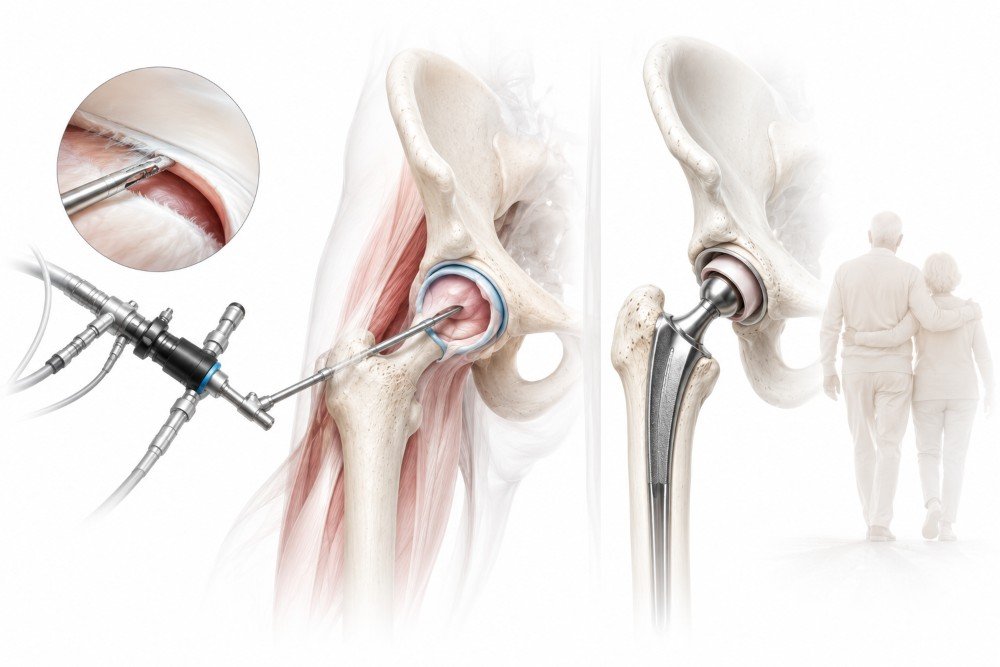
Surgery becomes relevant when conservative care has had a genuine trial without adequate result, or when the structural problem will not respond to anything else.
Hip arthroscopy addresses labral tears and impingement in suitable candidates through small incisions and a camera, reshaping bone where the anatomy is driving the friction and repairing the labrum where it can be salvaged.
Total hip replacement is reserved for advanced arthritis, and the durability picture here has changed substantially in the last few years. The widely quoted 2019 analysis, based largely on implants fitted before 2003, concluded that roughly 58 percent of hip replacements last twenty-five years. A much larger 2026 study drawing on nearly two million procedures across eight national joint registries, and looking specifically at modern bearing surfaces, reported survivorship above 93 percent at twenty years, with predicted survival above 92 percent at both twenty-five and thirty years.
If you were quoted the older figure, it is worth knowing that it described older hardware.
Red Flags That Change The Timeline
Most hip pain can wait for an ordinary appointment. Some cannot.
Seek urgent care if the pain follows a significant fall, if the leg is deformed or badly bruised, if you cannot bear weight or move the hip at all, or if hip pain arrives together with fever or a rash. Sudden hip pain in someone with sickle cell disease or a history of long-term steroid use also warrants prompt attention rather than a wait-and-see approach, because both raise the possibility of osteonecrosis.
These are not the situations this article is about, and they should not be managed by preparation and patience.
Closing Thoughts
The hip is the joint most likely to lie about its whereabouts. It sends pain to the groin, the thigh, the buttock, and the knee, and in the case of the buttock it does so more often than most people, including some clinicians, expect.
So before you see anyone, work out precisely where the pain sits, and be ready to say so. Note what provokes it. Note whether a long stretch of sitting makes it worse. Note whether anything catches or clicks. Note what you have stopped doing.
Then have it examined properly, by someone who will move the joint, test the spine, and treat the scan as evidence rather than as an answer. The location of your pain is the beginning of that conversation. It was never going to be the end of it.
Medical Disclaimer
This article is general information and not medical advice. It cannot diagnose your pain, and individual cases vary considerably. Severe hip pain that follows a significant fall, prevents you from bearing weight, leaves the leg deformed, or arrives alongside fever needs prompt medical attention rather than a considered wait. Speak with a licensed healthcare provider about your own symptoms before acting on anything described here.
References
- Lesher JM, Dreyfuss P, Hager N, Kaplan M, Furman M. Hip joint pain referral patterns: a descriptive study. Pain Medicine. 2008;9(1):22-25. https://doi.org/10.1111/j.1526-4637.2006.00153.x (PMID: 18254763)
- Khan AM, McLoughlin E, Giannakas K, Hutchinson C, Andrew JG. Hip osteoarthritis: where is the pain? Annals of the Royal College of Surgeons of England. 2004;86(2):119-121. https://doi.org/10.1308/003588404322827518 (PMID: 15005931; PMCID: PMC1964166)
- Dibra FF, Prieto HA, Gray CF, Parvataneni HK. Don’t forget the hip! Hip arthritis masquerading as knee pain. Arthroplasty Today. 2017;4(1):118-124. https://doi.org/10.1016/j.artd.2017.06.008 (PMID: 29560406; PMCID: PMC5859208)
- Griffin DR, Dickenson EJ, O’Donnell J, et al. The Warwick Agreement on femoroacetabular impingement syndrome (FAI syndrome): an international consensus statement. British Journal of Sports Medicine. 2016;50(19):1169-1176. https://doi.org/10.1136/bjsports-2016-096743 (PMID: 27629403)
- Evans JT, Evans JP, Walker RW, Blom AW, Whitehouse MR, Sayers A. How long does a hip replacement last? A systematic review and meta-analysis of case series and national registry reports with more than 15 years of follow-up. The Lancet. 2019;393(10172):647-654. https://doi.org/10.1016/S0140-6736(18)31665-9 (PMID: 30782340; PMCID: PMC6376618)
- Pentland V, Thompson Z, Dayimu A, et al. Survivorship of modern total hip replacement to 30 years: systematic review, meta-analysis, and extrapolation of global joint registry data. The Lancet. 2026;407(10531):855-866. https://doi.org/10.1016/S0140-6736(25)02305-0 (PMID: 41763743)