
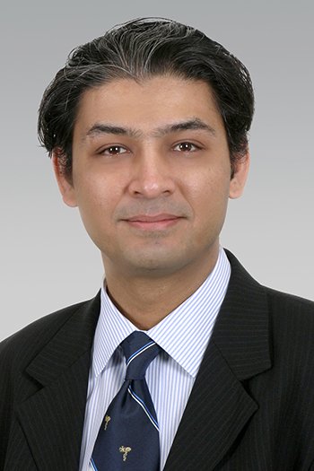
Dr. Adnan H. Siddiqui Cerebrovascular Neurosurgeon and Stroke Care
Dr. Adnan H. Siddiqui is a board-certified neurosurgeon whose clinical work focuses on cerebrovascular disorders, neuroendovascular treatment, stroke care, brain aneurysms, arteriovenous malformations, and complex conditions affecting blood vessels in the brain and spinal cord.
His practice combines open microsurgery with minimally invasive procedures performed through blood vessels. This allows treatment options to be considered according to the location of the problem, the patient’s symptoms, the risk of bleeding or stroke, and the expected benefits and limitations of each approach.
Dr. Siddiqui is a University at Buffalo Distinguished Professor and vice chairman of neurosurgery. He also holds leadership roles in neurovascular research, stroke services, specialist training, and medical technology development.
About Dr. Adnan H. Siddiqui
Dr. Siddiqui is a dual-trained cerebrovascular and neuroendovascular surgeon. His work centres on disorders affecting the arteries and veins that supply the brain, neck, face, and spinal cord.
Patients may be referred to him after imaging identifies an aneurysm, narrowed artery, blocked blood vessel, vascular malformation, fistula, or another complex abnormality. He may also evaluate people who have experienced an acute stroke or need another opinion about a proposed brain or blood-vessel procedure.
His care fits within both neurology and nervous system services and specialised surgical care. Treatment decisions in this field may involve neurologists, neurosurgeons, neuroradiologists, intensive-care physicians, rehabilitation professionals, and other specialists.
A consultation does not automatically mean that an operation will be recommended. Some vascular abnormalities can be monitored, while others may require medication, endovascular treatment, open surgery, radiosurgery, or a planned combination of approaches.
Professional Background and Leadership
Dr. Siddiqui joined University at Buffalo Neurosurgery in 2006. He is a Distinguished Professor and vice chairman in the Department of Neurosurgery at the Jacobs School of Medicine and Biomedical Sciences.
His current professional responsibilities include leadership in neuroendovascular education, neurosurgical research, stroke services, and vascular technology development. He serves as chief executive officer and chief medical officer of the Jacobs Institute and directs the Canon Stroke and Vascular Research Center.
He also directs a neuroendovascular fellowship programme and the neurosurgical stroke service connected with the Gates Vascular Institute. These roles combine clinical treatment with research, physician education, clinical trials, and the assessment of new devices used in vascular and neurological care.
Dr. Siddiqui has contributed to hundreds of peer-reviewed publications, book chapters, medical presentations, and national or international clinical trials. His research interests include stroke, brain aneurysms, vascular imaging, blood-flow modelling, medical devices, and techniques used in microsurgical and endovascular treatment.
Education and Specialist Training
| Training Area | Institution | Details |
|---|---|---|
| Medical education | Aga Khan University | Doctor of Medicine |
| Graduate study | University of Rochester | Neuroscience research and PhD training |
| Surgical internship | SUNY Upstate Medical University | Surgical internship |
| Neurosurgical residency | SUNY Upstate Medical University | Neurological surgery |
| Additional residency experience | University of Rochester | Neurology |
| Paediatric experience | Children’s Hospital Boston | Paediatric neurosurgery |
| Fellowship training | Thomas Jefferson University | Cerebrovascular surgery, interventional neuroradiology and neurocritical care |
Dr. Siddiqui’s training covers both open neurosurgery and catheter-based neuroendovascular care. Open microsurgery reaches the affected area through a surgical opening, while endovascular treatment uses thin catheters guided through blood vessels.
This combined training can be useful when more than one treatment method is medically reasonable. It allows the clinical team to compare options instead of approaching every condition through a single technique. His official professional profiles also list board certification in neurological surgery.
Board Certification and Professional Credentials
Dr. Siddiqui’s listed professional credentials include:
- MD, meaning Doctor of Medicine
- PhD, reflecting advanced research training in neuroscience
- FACS, meaning Fellow of the American College of Surgeons
- FAHA, meaning Fellow of the American Heart Association
- FAANS, meaning Fellow of the American Association of Neurological Surgeons
- Board certification in neurological surgery
Medical licensing, board certification, academic appointments, and professional fellowships represent different qualifications. Patients who need the latest credential information can confirm it through the relevant medical licensing and certification organisations.
Main Areas of Cerebrovascular Care
Brain Aneurysms
A cerebral or brain aneurysm is a weakened area in the wall of an artery that becomes enlarged or balloon-shaped. Some aneurysms are found during imaging performed for an unrelated reason, while others cause symptoms or are detected after bleeding.
The National Institute of Neurological Disorders and Stroke provides patient information about cerebral aneurysms, including diagnosis, monitoring, clipping, and endovascular treatment.
Dr. Siddiqui’s documented clinical work includes microsurgical and endovascular management of brain aneurysms. Depending on the individual case, options may include:
- Monitoring with repeat imaging
- Management of blood pressure and other risk factors
- Microsurgical clipping
- Endovascular coiling
- Flow-diverting devices
- Intrasaccular devices placed inside an aneurysm
- Treatment following aneurysm rupture
- Combined treatment for unusually complex aneurysms
The decision depends on the aneurysm’s size, shape, location, growth, rupture status, nearby blood vessels, and the patient’s age and general health.
Arteriovenous Malformations
An arteriovenous malformation, commonly called an AVM, is an abnormal connection between arteries and veins. It may occur in the brain or spinal cord and can interfere with normal blood flow.
Some AVMs are discovered after a seizure, headache, neurological change, or brain bleed. Others are found while investigating a different medical concern.
Patients can review information about arteriovenous malformations from NINDS before discussing their individual imaging and treatment choices with a specialist.
Possible treatment approaches may include:
- Careful monitoring
- Endovascular embolisation
- Open microsurgical removal
- Stereotactic radiosurgery
- Treatment divided into more than one stage
- A combination of embolisation, surgery, and radiosurgery
An AVM may be too complicated for one treatment method alone. The clinical team must consider its size, position, blood supply, drainage pattern, history of bleeding, and relationship to important areas of the brain.
Dural and Cavernous Fistulas
A fistula is an abnormal connection that allows blood to pass directly between an artery and a vein.
A dural arteriovenous fistula develops within the protective covering around the brain or spinal cord. A carotid-cavernous fistula affects blood vessels behind the eye and may cause eye redness, swelling, vision changes, a pulsing sound, or pressure around the eye.
Dr. Siddiqui’s listed interests include dural, cavernous, and spinal fistulas. Treatment may involve catheter angiography, embolisation, surgery, radiosurgery, or observation, depending on the blood-flow pattern and risk of complications.
Acute Ischemic Stroke
An ischemic stroke happens when a blood vessel supplying the brain becomes blocked. Brain tissue can be damaged when blood flow is not restored quickly.
Stroke treatment is time-sensitive. Patients with sudden facial weakness, arm weakness, speech difficulty, confusion, vision loss, severe imbalance, or another abrupt neurological change need emergency assessment rather than a routine clinic appointment.
Dr. Siddiqui’s work includes endovascular management of acute ischemic stroke. This may involve mechanical thrombectomy, a procedure in which specialists guide a catheter through an artery and use a device to remove a clot from a large brain blood vessel.
NINDS explains how specialists assess and treat stroke, including the role of brain imaging, clot-dissolving medication, thrombectomy, and treatment for bleeding in the brain.
Intracranial and Extracranial Vascular Occlusive Disease
Vascular occlusive disease involves narrowing or blockage of arteries that carry blood to the brain. The affected vessel may be inside the skull or within the neck.
Possible causes include plaque buildup, blood clots, vessel injury, inflammation, or less common blood-vessel disorders. Symptoms may include transient ischemic attacks, strokes, weakness, speech changes, vision problems, or episodes of reduced brain blood flow.
Assessment may involve:
- CT angiography
- MR angiography
- Cerebral angiography
- Ultrasound of neck arteries
- Blood-flow studies
- Review of previous strokes or transient symptoms
- Evaluation of blood pressure, cholesterol, diabetes, and smoking history
Treatment may involve medication and risk-factor management, while selected patients may require angioplasty, stenting, bypass surgery, or another procedure.
Moyamoya Disease
Moyamoya disease is a rare condition in which major arteries supplying the brain become progressively narrowed. The brain may form small alternative vessels to compensate, but these vessels may not provide enough reliable blood flow.
Patients may experience transient ischemic attacks, strokes, headaches, seizures, weakness, or cognitive changes. Treatment decisions depend on symptoms, brain-blood-flow testing, and the severity of arterial narrowing.
In 2025, Dr. Siddiqui performed investigational robotic-assisted intracranial microsurgical procedures during an early feasibility study involving adults with moyamoya disease. These cases were part of a clinical study and should not be understood as established routine treatment for every patient.
Skull Base and Brain Tumour Care
Skull base tumours develop near the lower part of the skull, where important nerves and blood vessels are closely packed together. Treatment may require cooperation between neurosurgery, ear and throat surgery, radiology, oncology, and other specialties.
Dr. Siddiqui’s clinical interests include microsurgical removal of selected skull base tumours and embolisation of certain head, neck, and brain tumours.
Embolisation blocks or reduces blood flow to abnormal tissue. It may be performed before surgery when reducing a tumour’s blood supply could make the operation more manageable.
The National Cancer Institute provides patient information about brain tumours and their treatment. The correct treatment depends on the tumour type, size, position, growth pattern, symptoms, and relationship to nearby structures.
Conditions Dr. Siddiqui May Evaluate
Dr. Siddiqui’s verified clinical interests include assessment and treatment planning for:
- Cerebral aneurysms
- Ruptured brain aneurysms
- Arteriovenous malformations
- Dural arteriovenous fistulas
- Carotid-cavernous fistulas
- Spinal vascular fistulas
- Acute ischemic stroke
- Intracranial arterial narrowing
- Extracranial vascular occlusive disease
- Moyamoya disease
- Facial vascular malformations
- Severe or difficult-to-control nosebleeds requiring embolisation
- Brain and head tumour embolisation
- Skull base tumours
- Pituitary tumours in selected cases
- Arachnoid cysts
- Hydrocephalus requiring specialist assessment
- Complex cerebrovascular conditions needing a second opinion
This list does not mean that every patient with one of these diagnoses requires surgery. Symptoms, imaging, medical history, previous treatment, and the natural behaviour of the condition must all be considered.
Procedures Connected With His Practice
Cerebral Angiography
Cerebral angiography provides detailed images of blood vessels in the brain, head, and neck. A thin catheter is guided through an artery before contrast material is used to show blood flow.
Angiography may help identify:
- An aneurysm’s exact shape and location
- The vessels supplying an AVM
- Abnormal connections between arteries and veins
- Narrowed or blocked arteries
- Blood-vessel changes near a tumour
- Whether an earlier procedure remains effective
NINDS provides information about neurological diagnostic tests and procedures, including cerebral angiography, CT, MRI, and other assessments.
Endovascular Embolisation
Embolisation uses a catheter to place material into an abnormal blood vessel or area of unwanted blood flow.
Depending on the condition, embolisation may use coils, liquid embolic materials, particles, plugs, or another specialised device. It may be the main treatment or one stage of a larger plan involving surgery or radiosurgery.
Aneurysm Coiling and Flow Diversion
Coiling places small devices inside an aneurysm to reduce blood entry and encourage clotting within the weakened area.
Flow diversion places a specialised device within the parent artery to redirect blood away from the aneurysm. The right option depends on the aneurysm’s anatomy and whether important branches arise nearby.
Microsurgical Aneurysm Clipping
Clipping is an open surgical procedure in which a small clip is placed across the neck of an aneurysm to separate it from normal blood circulation.
Clipping may be considered when the aneurysm’s location or shape makes open surgery suitable, when nearby blood vessels need direct visual protection, or when another treatment is less appropriate.
Mechanical Thrombectomy
Mechanical thrombectomy is an emergency stroke procedure used for selected large-vessel blockages. A catheter-based device is guided to the clot so blood flow can be restored.
Eligibility depends on when symptoms began, which artery is blocked, brain-imaging findings, the amount of brain tissue already affected, and the patient’s overall condition.
Stereotactic Radiosurgery
Stereotactic radiosurgery directs concentrated radiation towards a precisely defined target without making a traditional surgical opening.
It may be considered for selected AVMs and other abnormalities. The effect develops gradually, so it may not provide immediate protection from bleeding.
Tumour Embolisation
Tumour embolisation reduces blood flow to selected tumours before another treatment. It may help with surgical planning, but it is not appropriate for every tumour.
The clinical team must consider whether the tumour can be safely reached through the blood vessels and whether reducing its blood supply is likely to help.
How Cerebrovascular Conditions Are Assessed
A specialist assessment usually begins with a detailed review of the event or symptoms that led to the referral.
Patients may be asked about:
- Sudden or severe headaches
- Previous strokes or transient ischemic attacks
- Weakness or numbness
- Speech or vision changes
- Seizures
- Balance problems
- Previous brain bleeding
- Family history of aneurysms
- Blood pressure
- Smoking history
- Earlier operations or endovascular procedures
- Blood-thinning medication
- Kidney problems or contrast allergies
The medical team may review:
- CT scans
- MRI scans
- CT angiography
- MR angiography
- Cerebral angiography
- Ultrasound studies
- Perfusion or blood-flow imaging
- Earlier surgical reports
- Hospital discharge records
- Medication history
Imaging should be interpreted alongside symptoms and examination findings. A vascular abnormality found on a scan does not always explain every symptom a patient experiences.
Who May Benefit From Seeing Dr. Siddiqui
A specialist consultation may be useful for patients who:
- Have been diagnosed with a brain aneurysm
- Need treatment after aneurysm rupture
- Have an AVM or vascular fistula
- Have experienced an ischemic or haemorrhagic stroke
- Have narrowing or blockage in a brain artery
- Need an opinion about endovascular treatment
- Have been advised to consider aneurysm clipping
- Need a second opinion about a complex vascular condition
- Have had an earlier procedure that requires follow-up
- Have a skull base tumour involving important blood vessels
- Need tumour embolisation before surgery
- Have several possible treatment options and need help comparing them
- Have been told that their condition requires a multidisciplinary team
Patients comparing professionals can also use the Doctiplus doctor and specialist directory to review other relevant care options.
What Patients Can Expect During an Appointment
The first appointment may focus on understanding the diagnosis and deciding whether more testing is needed.
Dr. Siddiqui or the clinical team may discuss:
- How the condition was discovered
- Whether symptoms have occurred
- The risk of bleeding or stroke
- The size and location of the abnormality
- Whether it has changed on repeat imaging
- Treatments already attempted
- The benefits and risks of monitoring
- Open surgical options
- Catheter-based options
- Radiosurgical treatment
- The possibility of staged or combined care
- Follow-up imaging requirements
Patients should ask for medical terms and imaging findings to be explained in plain language. Understanding why one option is being recommended is as important as knowing the name of the procedure.
Preparing for the Consultation
Patients may find it helpful to bring:
- CT, MRI, and angiography images
- Written radiology reports
- Hospital discharge documents
- Previous operative reports
- A list of current medicines
- Details of blood thinners or antiplatelet medicines
- Medication and contrast allergies
- A record of earlier strokes or transient symptoms
- Blood pressure history
- Relevant family medical history
- A timeline of headaches or neurological changes
- Insurance and referral documents
- A written list of questions
Patients should not stop aspirin, anticoagulants, or other prescribed medicines before an appointment unless the prescribing clinician provides specific instructions.
Questions to Ask During the Consultation
Useful questions may include:
- What does my scan show?
- Is the abnormality causing my symptoms?
- What is the risk of monitoring it?
- What factors increase the risk of bleeding or stroke?
- Do I need more imaging?
- Is open surgery or endovascular treatment more suitable?
- Could radiosurgery be considered?
- Why is this particular procedure recommended?
- Are there reasonable alternatives?
- Could treatment require more than one stage?
- What are the main risks?
- How much experience does the treating team have with this condition?
- Which medicines will I need before and after treatment?
- How long may hospital care and recovery take?
- How often will follow-up imaging be required?
- Would a second opinion change the treatment plan?
- Which symptoms require emergency care?
Second Opinions for Complex Brain-Vessel Conditions
A second opinion can be especially useful when an aneurysm, AVM, fistula, or narrowed artery could be treated in several different ways.
Another review may help when:
- The diagnosis is uncertain
- The condition is uncommon
- A major operation has been recommended
- Both open and endovascular treatments are possible
- Previous treatment was incomplete
- The abnormality has returned
- The estimated risks are difficult to understand
- The patient wants another specialist to review the original imaging
A second opinion may confirm the first recommendation or identify another reasonable approach. It does not necessarily mean that the original medical team made an incorrect decision.
Recovery and Follow-Up Care
Recovery varies according to the diagnosis and procedure. A diagnostic angiogram may require a relatively short recovery, while treatment for a ruptured aneurysm, major stroke, AVM, or skull base tumour can require extended hospital care and rehabilitation.
Follow-up may include:
- Neurological examinations
- Wound or catheter-entry checks
- Medication review
- CT or MRI imaging
- Repeat angiography
- Blood pressure management
- Stroke-prevention planning
- Physical therapy
- Occupational therapy
- Speech and language therapy
- Cognitive rehabilitation
- Return-to-work guidance
- Long-term monitoring for recurrence or vessel changes
Patients should follow the individual instructions provided by their treatment team. Recovery expectations can differ substantially even when two people undergo procedures with similar names.
Research and Surgical Innovation
Dr. Siddiqui’s academic work includes clinical trials, medical-device evaluation, imaging, stroke research, aneurysm research, and collaboration with engineers and scientists.
In 2025, he used an investigational robotic microsurgical system during the first reported robotic-assisted intracranial microsurgical procedures in an early feasibility study. The cases involved indirect bypass surgery for adults with moyamoya disease.
Because the technology was being evaluated in a regulated clinical study, it should not be presented as a standard option available to every neurosurgical patient. Research procedures require specific eligibility criteria, consent, oversight, and follow-up.
Practice and Appointment Information
Dr. Siddiqui sees patients through University at Buffalo Neurosurgery. His official clinical profile lists the Comprehensive Neuroscience Center as an appointment location:
Comprehensive Neuroscience Center
40 George Karl Boulevard, Suite 100
Williamsville, New York 14221
Phone: 716-218-1000
Clinic location, appointment availability, referral rules, and the facility used for a procedure can vary according to the condition and required treatment. Patients should confirm the current details when scheduling.
Insurance and Referral Preparation
Before booking, patients may wish to ask:
- Is a physician referral required?
- Is Dr. Siddiqui included in my insurance network?
- Does the clinic need imaging before accepting the referral?
- Should original image files be uploaded or brought on a disc?
- Does insurance require prior authorisation?
- Are diagnostic angiography and treatment approved separately?
- Which hospital would provide care?
- Are second-opinion consultations covered?
- Are remote imaging reviews available?
- Will rehabilitation services need separate approval?
Insurance approval for an office appointment may not automatically cover imaging, hospital care, medical devices, surgery, rehabilitation, or follow-up procedures.
When Symptoms Require Emergency Care
A routine appointment is not suitable for possible stroke, aneurysm rupture, or sudden bleeding in the brain.
Seek emergency medical care for:
- Sudden weakness or numbness on one side
- Facial drooping
- New speech difficulty
- Sudden confusion
- Sudden loss of vision
- Severe difficulty walking or maintaining balance
- A sudden, extremely severe headache
- Collapse or loss of consciousness
- A first seizure
- Repeated vomiting with a severe headache
- Sudden neck stiffness with neurological symptoms
- Rapidly worsening symptoms after a known aneurysm or AVM diagnosis
Do not drive yourself when stroke or brain bleeding is suspected. Contact emergency medical services so assessment and treatment can begin as quickly as possible.
Profile Accuracy
This profile has been prepared using current publicly available professional, academic, clinical, and institutional information.
Readers can review the Doctiplus editorial policy to understand how patient-focused health content is prepared and maintained.
Doctiplus also provides a collection of trusted healthcare information resources used to support responsible explanations of diseases, tests, and treatments.



Reviews
There are no reviews yet.