

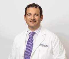
Dr. Asif Akhtar Interventional Cardiologist
Dr. Asif Akhtar is an interventional cardiologist providing diagnostic, preventive, and procedural care for adults with heart and blood-vessel conditions.
His clinical work includes coronary artery disease, chest pain, high blood pressure, high cholesterol, heart failure, abnormal heart rhythms, heart-valve disorders, circulation problems, and cardiovascular risk assessment.
As an interventional cardiologist, Dr. Akhtar may also evaluate patients who require cardiac catheterisation, coronary angiography, angioplasty, or stent treatment. These procedures allow a cardiologist to examine or treat narrowed blood vessels through small catheter-based access rather than traditional open surgery.
Dr. Akhtar practises at Interventional Cardiology Associates in Houston, Texas. Patients may visit him because of a new heart symptom, an abnormal diagnostic test, an existing cardiovascular diagnosis, or a recommendation for an interventional procedure.
Patients comparing cardiology specialists may also read about Dr. Azadeh Beheshtian’s interventional cardiology care.
Doctor Information
Name: Dr. Asif Akhtar, MD
Specialty: Interventional Cardiology
Additional Areas: Cardiovascular disease and internal medicine
Board Certification: Cardiovascular Disease
Medical School: Dow Medical College
Internship: Internal Medicine, Henry Ford Hospital
Residency: Internal Medicine, Henry Ford Hospital
Fellowship Training: Cardiovascular medicine through Henry Ford Hospital, according to current professional listings
Practice: Interventional Cardiology Associates
Primary Location: Houston, Texas
Office Area: Park Row in West Houston
Hospital Affiliations: Current profiles list HCA Houston Healthcare Medical Center and Houston Methodist facilities
Languages Listed: English and Arabic; some directories also list Spanish
NPI Number: 1801900352
Accepting New Patients: Yes, according to current provider listings
About Dr. Asif Akhtar
Dr. Akhtar completed his medical education at Dow Medical College before continuing postgraduate training in the United States.
His professional records list internship, residency, and cardiovascular fellowship education through Henry Ford Hospital in Detroit, Michigan.
Internal medicine training prepared him to care for adult medical conditions involving several body systems. He then continued into cardiovascular medicine, focusing on diseases affecting the heart and blood vessels.
Interventional cardiology adds specialised catheter-based training. An interventional cardiologist can use narrow tubes, imaging, balloons, stents, and other devices to diagnose or treat selected cardiovascular conditions.
A patient may visit Dr. Akhtar because of chest discomfort, shortness of breath, palpitations, dizziness, swelling, an abnormal heart test, or an established condition requiring continuing monitoring.
His evaluation may include a medical history, physical examination, electrocardiogram, ultrasound, stress testing, vascular testing, blood work, or more advanced cardiac imaging.
Interventional Cardiology Services
Dr. Akhtar may evaluate and manage concerns such as:
- Coronary artery disease
- Chest pain and angina
- Previous heart attack
- Acute coronary syndromes
- High blood pressure
- High cholesterol
- Atherosclerosis
- Heart failure
- Cardiomyopathy
- Abnormal heart rhythms
- Atrial fibrillation
- Heart-valve disorders
- Fainting or near-fainting
- Peripheral artery disease
- Swelling associated with cardiovascular conditions
- Abnormal cardiovascular test results
His procedural work may include or coordinate:
- Cardiac catheterisation
- Coronary angiography
- Angioplasty
- Coronary stent placement
- Cardiovascular stress testing
- Echocardiography
- Nuclear cardiac imaging
- Vascular ultrasound
- Structural heart evaluation
The exact services available should be confirmed directly with the practice because procedures may be performed at different hospitals or cardiovascular facilities.
Coronary Artery Disease
Coronary artery disease develops when plaque builds up within the arteries that supply blood to the heart muscle.
Plaque may gradually narrow the artery and reduce blood flow. A plaque can also rupture, leading to a blood clot and possible heart attack.
Possible symptoms include:
- Chest pressure
- Chest tightness
- Discomfort during physical activity
- Shortness of breath
- Reduced exercise tolerance
- Pain extending toward the arm, shoulder, jaw, back, or upper abdomen
- Unusual tiredness
- Nausea or sweating during exertion
Some patients have few symptoms even when significant narrowing is present.
Dr. Akhtar may assess symptoms, risk factors, electrocardiogram findings, stress-test results, and cardiac imaging before deciding whether further testing is necessary.
Treatment may involve medicine and cardiovascular risk management. Cardiac catheterisation or an interventional procedure may be considered when symptoms, test findings, or clinical risks suggest significant artery narrowing.
Chest Pain Evaluation
Chest pain does not always originate from the heart, but cardiovascular causes need careful consideration.
Dr. Akhtar may ask:
- When the discomfort began
- Where it is located
- Whether it feels like pressure, burning, tightness, or pain
- Whether physical activity brings it on
- Whether rest improves it
- How long each episode lasts
- Whether breathing affects it
- Whether it travels to another area
- Whether sweating, nausea, or shortness of breath occurs
- Which cardiovascular risks are present
Evaluation may include an electrocardiogram, blood tests, echocardiogram, stress testing, CT imaging, or coronary angiography.
The correct test depends on the symptom pattern, age, previous heart history, and overall cardiovascular risk.
Angina
Angina is chest discomfort caused by insufficient blood flow to the heart muscle.
Stable angina often follows a recognisable pattern. It may occur during exercise, emotional stress, or another activity that makes the heart work harder.
Unstable angina may be new, more severe, more frequent, or present at rest. It may indicate an increased risk of a heart attack.
Dr. Akhtar may review:
- Symptom frequency
- Exercise tolerance
- Medicine response
- Previous stress testing
- Coronary imaging
- Existing stents or bypass surgery
- Blood pressure and cholesterol
- Diabetes
- Tobacco exposure
Treatment may include medicine, risk-factor control, cardiac catheterisation, or coronary intervention.
Cardiac Catheterisation
Cardiac catheterisation uses a narrow flexible tube inserted into a blood vessel and guided toward the heart.
The procedure may help measure pressure, assess heart function, and identify narrowing in the coronary arteries.
It may be considered when:
- Chest symptoms suggest reduced heart blood flow
- A stress test is abnormal
- Symptoms continue despite treatment
- A heart attack is suspected
- Previous stents require assessment
- Structural heart disease needs further evaluation
The catheter may enter through an artery in the wrist or groin.
Contrast material is usually injected so the coronary arteries can be viewed on X-ray imaging.
A diagnostic catheterisation may show that medicine is the best approach. In other cases, angioplasty or stent placement may be performed during the same procedure.
Coronary Angiography
Coronary angiography creates moving X-ray images of blood flow through the heart arteries.
It may identify:
- The location of narrowing
- The severity of blockage
- The number of affected arteries
- Blood flow through previous stents
- Whether bypass grafts remain open
- Whether an interventional procedure may help
The findings are interpreted alongside symptoms, heart function, age, kidney health, bleeding risks, and other medical conditions.
Not every visible narrowing requires a stent. Some conditions are managed more effectively with medicine, cardiovascular risk reduction, or bypass surgery.
Angioplasty
Angioplasty opens a narrowed artery with a small balloon attached to a catheter.
The balloon is positioned within the narrowed area and expanded to improve blood flow.
Angioplasty is commonly combined with stent placement.
It may be used during treatment of:
- A heart attack
- Severe or continuing angina
- Significant coronary narrowing
- Reduced blood flow confirmed by testing
- Selected problems involving an earlier stent
The expected benefit depends on the location and severity of the blockage, the number of affected arteries, heart function, and the patient’s general health.
Coronary Stent Placement
A coronary stent is a small mesh tube used to support an artery after it has been opened.
Most current coronary stents release medicine that helps reduce unwanted tissue growth inside the treated area.
After stent placement, patients commonly need antiplatelet medicines to reduce the risk of a clot forming within the stent.
Patients should understand:
- Which artery was treated
- How many stents were placed
- Which medicines are required
- How long antiplatelet treatment may continue
- Which activities are temporarily restricted
- When follow-up is needed
- Which symptoms should be reported to the medical team
Antiplatelet treatment should not be stopped without guidance from the cardiologist or another responsible physician. Stopping it too soon may create a serious risk.
Heart Attack Care
A heart attack develops when blood flow through a coronary artery becomes severely reduced or blocked.
Treatment may include urgent cardiac catheterisation, angioplasty, and stent placement.
After hospital treatment, Dr. Akhtar may help manage:
- Antiplatelet medicine
- Cholesterol-lowering treatment
- Blood pressure
- Heart rate
- Diabetes
- Heart function
- Cardiac rehabilitation
- Return to activity
- Follow-up testing
Recovery is not limited to the treated artery. Continuing care also focuses on reducing the likelihood of another cardiovascular event.
High Blood Pressure
High blood pressure may cause no noticeable symptoms while gradually affecting the heart, kidneys, brain, eyes, and blood vessels.
Dr. Akhtar may review:
- Office blood pressure readings
- Home measurements
- Current medicines
- Kidney function
- Heart structure
- Salt intake
- Physical activity
- Sleep quality
- Other cardiovascular risks
Long-term high blood pressure can contribute to:
- Thickening of the heart muscle
- Heart failure
- Coronary artery disease
- Stroke
- Kidney impairment
- Aortic disease
- Abnormal heart rhythms
Patients using a home monitor should record the date, time, reading, and whether medicine was taken before the measurement.
High Cholesterol
High cholesterol contributes to plaque formation within the arteries.
A cholesterol assessment may include:
- Total cholesterol
- LDL cholesterol
- HDL cholesterol
- Triglycerides
- Family history
- Diabetes
- Blood pressure
- Tobacco exposure
- Kidney health
- Previous cardiovascular disease
Treatment may involve nutrition changes, physical activity, medicine, or a combination of approaches.
Patients with established coronary artery disease often require more intensive cholesterol management than people receiving preventive care before cardiovascular disease develops.
The treatment target should be based on total cardiovascular risk rather than one number alone.
Atherosclerosis
Atherosclerosis is the buildup of plaque within arteries.
It can affect blood vessels supplying the:
- Heart
- Brain
- Legs
- Kidneys
- Other organs
The condition may remain silent for years.
When it becomes significant, symptoms depend on which artery is affected. Coronary disease may cause angina, while peripheral artery disease may cause leg discomfort during walking.
Dr. Akhtar may assess whether atherosclerosis is limited to one area or reflects a broader blood-vessel condition requiring comprehensive risk management.
Heart Failure
Heart failure means the heart is not pumping or filling as effectively as the body needs.
Possible symptoms include:
- Shortness of breath
- Swelling in the legs
- Sudden weight gain from fluid
- Difficulty lying flat
- Nighttime breathing problems
- Reduced exercise tolerance
- Tiredness
- Reduced appetite
Heart failure can develop because of coronary artery disease, previous heart attack, high blood pressure, valve disease, abnormal rhythms, or cardiomyopathy.
Dr. Akhtar may review heart imaging, laboratory results, weight patterns, kidney function, blood pressure, and medicine.
Treatment may include several medicines, fluid management, treatment of coronary disease, rhythm care, valve assessment, and coordination with a heart-failure specialist.
Cardiomyopathy
Cardiomyopathy describes disease affecting the heart muscle.
The heart may become enlarged, thickened, weakened, or unusually stiff.
Possible symptoms include:
- Shortness of breath
- Tiredness
- Swelling
- Palpitations
- Chest discomfort
- Dizziness
- Fainting
- Reduced exercise ability
Possible causes include inherited conditions, coronary artery disease, uncontrolled blood pressure, infection, toxins, medication, or another medical condition.
Evaluation may include echocardiography, electrocardiography, cardiac MRI, blood testing, rhythm monitoring, or coronary assessment.
Treatment depends on the type and cause.
Atrial Fibrillation
Atrial fibrillation is an abnormal heart rhythm arising in the upper chambers of the heart.
Symptoms may include:
- Palpitations
- Irregular heartbeat
- Shortness of breath
- Tiredness
- Dizziness
- Reduced exercise tolerance
- Chest discomfort
Some patients have no obvious symptoms.
Atrial fibrillation can increase the risk of stroke. Dr. Akhtar may assess whether blood-thinning medicine is appropriate based on age, medical history, bleeding risk, and other factors.
Treatment may also involve heart-rate control, rhythm treatment, cardioversion, or referral to an electrophysiologist.
Palpitations
Palpitations may feel like a racing, pounding, fluttering, skipped, or irregular heartbeat.
Possible causes include:
- Atrial fibrillation
- Premature heartbeats
- Other rhythm disorders
- Caffeine
- Medicine
- Thyroid disease
- Anemia
- Dehydration
- Stress
- Sleep loss
Testing may include an electrocardiogram, portable rhythm monitor, blood tests, or echocardiogram.
Because rhythm disturbances may occur intermittently, a short office electrocardiogram may not capture every episode.
Heart-Valve Disease
The heart contains valves that keep blood moving in the correct direction.
A valve may become narrowed or may fail to close completely.
Dr. Akhtar may evaluate conditions involving the:
- Aortic valve
- Mitral valve
- Tricuspid valve
- Pulmonary valve
Possible symptoms include:
- Shortness of breath
- Chest discomfort
- Tiredness
- Swelling
- Palpitations
- Dizziness
- Fainting
- Reduced exercise tolerance
Echocardiography is commonly used to assess valve structure, movement, narrowing, and leakage.
Treatment may involve continued monitoring, medicine, catheter-based intervention, or surgery.
Aortic Stenosis
Aortic stenosis occurs when the aortic valve becomes narrowed.
As narrowing progresses, the heart must work harder to move blood through the valve.
Possible symptoms include:
- Shortness of breath
- Chest pressure
- Dizziness
- Fainting
- Reduced exercise tolerance
- Tiredness
Severity is assessed through echocardiography and the patient’s symptoms.
Patients with advanced disease may need evaluation for surgical or transcatheter valve replacement.
The decision depends on valve severity, age, general health, anatomy, and expected procedural benefit.
Mitral Valve Disease
The mitral valve controls blood flow between the left upper and lower chambers of the heart.
Problems may include narrowing, leakage, prolapse, or changes related to infection or heart-muscle disease.
Dr. Akhtar may monitor:
- Symptom development
- Valve severity
- Heart size
- Heart pumping function
- Lung pressure
- Abnormal rhythms
Some patients only need periodic echocardiograms. Others may require a catheter-based or surgical valve procedure.
Peripheral Artery Disease
Peripheral artery disease occurs when narrowed arteries reduce blood flow to the limbs, most commonly the legs.
Possible symptoms include:
- Calf or thigh discomfort while walking
- Foot pain at rest
- Slow-healing wounds
- Coldness in one foot
- Weak leg pulses
- Skin-colour changes
- Reduced walking distance
Risk factors include tobacco exposure, diabetes, high cholesterol, high blood pressure, and established coronary artery disease.
Evaluation may include pulse examination, ankle-brachial index testing, vascular ultrasound, CT angiography, or another circulation study.
Treatment may include medicine, supervised exercise, risk-factor control, wound care, or an interventional procedure.
Deep Vein Thrombosis
Deep vein thrombosis is a blood clot within a deep vein, commonly in the leg.
Possible symptoms include:
- One-sided leg swelling
- Pain or tenderness
- Warmth
- Skin-colour changes
- A feeling of heaviness
Evaluation commonly involves vascular ultrasound.
Treatment often includes blood-thinning medicine to prevent the clot from growing or travelling to the lungs.
The length of treatment depends on the clot’s location, possible cause, recurrence risk, bleeding risk, and other medical conditions.
Aortic Aneurysm
An aortic aneurysm is an abnormal enlargement of part of the body’s main artery.
It may involve the chest or abdomen and often causes no symptoms before it is identified through imaging.
Dr. Akhtar may help assess:
- Aneurysm size
- Rate of growth
- Blood pressure
- Tobacco exposure
- Family history
- Associated valve disease
- Need for repeat imaging
- Need for surgical or vascular consultation
Smaller aneurysms may be monitored. Larger, rapidly growing, or symptomatic aneurysms may require procedural evaluation.
Fainting and Syncope
Syncope is a temporary loss of consciousness caused by reduced blood flow to the brain.
Possible cardiovascular causes include:
- Abnormal heart rhythms
- Valve disease
- Blood pressure changes
- Reduced heart pumping
- Medication effects
- Problems with nervous-system control of blood pressure
Dr. Akhtar may review what happened before and after the event, whether warning symptoms occurred, and whether injury resulted.
Testing may include an electrocardiogram, rhythm monitor, echocardiogram, stress test, or tilt-table study.
Electrocardiogram
An electrocardiogram, commonly called an ECG or EKG, records the heart’s electrical activity.
It may show:
- Heart rate
- Heart rhythm
- Signs of a previous heart attack
- Changes suggesting reduced blood flow
- Electrical-conduction problems
- Heart-muscle strain
- Medicine-related changes
The test is quick and does not send electricity into the body.
A normal electrocardiogram does not rule out every heart condition, particularly when symptoms occur intermittently.
Echocardiography
An echocardiogram uses ultrasound to create moving images of the heart.
It may assess:
- Heart pumping strength
- Chamber size
- Valve function
- Heart-wall movement
- Fluid around the heart
- Blood-flow patterns
- Pressure within the heart and lungs
The test can support evaluation of heart failure, murmurs, valve disorders, cardiomyopathy, and symptoms such as shortness of breath.
Cardiovascular Stress Testing
A stress test assesses how the heart responds when it works harder.
Exercise testing may involve walking on a treadmill while heart rhythm, symptoms, and blood pressure are monitored.
Patients who cannot exercise adequately may receive medicine that temporarily creates a stress-like effect.
Stress testing may be combined with:
- Electrocardiography
- Echocardiography
- Nuclear imaging
The test may help estimate whether the heart receives enough blood during activity.
An abnormal result does not by itself determine treatment. Findings must be interpreted with symptoms, medical history, and other test results.
Nuclear Cardiac Imaging
Nuclear cardiac imaging uses a small amount of radioactive tracer to assess blood flow to the heart muscle.
Images are usually collected during rest and stress.
The test may help identify:
- Reduced blood flow
- Previous heart-muscle damage
- Areas at risk during activity
- Heart pumping function
Results can help determine whether medicine, cardiac catheterisation, or another investigation should be considered.
Preparing for a Cardiology Appointment
Patients can prepare by bringing:
- A current medicine list
- Previous cardiology records
- Electrocardiogram results
- Echocardiogram reports
- Stress-test results
- Cardiac catheterisation reports
- Stent information
- Hospital discharge documents
- Blood pressure records
- Recent blood tests
- A list of medicine allergies
- Questions about diagnosis or treatment
Patients should explain:
- When symptoms began
- What triggers them
- How long they last
- Whether physical activity makes them worse
- Whether symptoms travel to another area
- Whether swelling or weight changes have occurred
- Which treatments have already been attempted
Actual imaging or procedure records may provide information beyond a short written summary.
Preparing for Cardiac Catheterisation
Preparation may involve:
- Blood tests
- Kidney-function assessment
- Medicine review
- Allergy review
- Instructions about eating and drinking
- Diabetes-treatment instructions
- Transportation arrangements
- Planning for help after discharge
Patients should tell the procedural team about:
- Blood thinners
- Diabetes medicine
- Insulin
- Contrast-material reactions
- Kidney disease
- Bleeding problems
- Previous catheterisation
- Existing stents
- Other medical conditions
Medicine should not be stopped unless the cardiology or prescribing team provides clear instructions.
Recovery After a Catheter Procedure
Recovery depends on the procedure, access site, medical health, and whether angioplasty or stent placement was performed.
Instructions may cover:
- Wrist or groin care
- Temporary lifting restrictions
- Walking
- Driving
- Hydration
- Medicine use
- Antiplatelet treatment
- Return to work
- Follow-up visits
Patients should follow the written discharge instructions even when they feel well.
The artery access site and treated blood vessel need time to heal.
Cardiac Rehabilitation
Cardiac rehabilitation is a supervised programme that may be recommended after a heart attack, coronary intervention, or another qualifying heart condition.
It may include:
- Monitored exercise
- Education about heart medicines
- Nutrition support
- Blood pressure management
- Diabetes management
- Tobacco-cessation support
- Guidance for returning to activity
The programme helps patients rebuild confidence and physical ability while cardiovascular risks are monitored.
Coordinated Cardiovascular Care
Heart and vascular conditions may require cooperation between several healthcare professionals.
Dr. Akhtar may coordinate with:
- Primary care physicians
- General cardiologists
- Cardiac electrophysiologists
- Heart-failure specialists
- Cardiothoracic surgeons
- Vascular surgeons
- Kidney specialists
- Endocrinologists
- Cardiac rehabilitation teams
Coordination is particularly important when a patient has coronary artery disease along with diabetes, kidney disease, heart failure, valve disease, or an abnormal heart rhythm.
Patients seeking broader preventive cardiology information may also review Dr. Gargi Bajpayee’s cardiovascular care profile and Dr. David Bolon’s preventive cardiology profile.
Patient-Centered Interventional Cardiology
Dr. Akhtar provides cardiovascular care based on each patient’s symptoms, examination, diagnostic results, previous procedures, medical risks, and treatment goals.
His services may include cardiovascular risk assessment, chest-pain evaluation, blood pressure and cholesterol management, heart-failure care, rhythm assessment, cardiac imaging, coronary angiography, angioplasty, and stent placement.
Patients should confirm current appointment availability, accepted insurance plans, hospital participation, procedure locations, telehealth options, and referral requirements directly with Interventional Cardiology Associates.



Reviews
There are no reviews yet.