

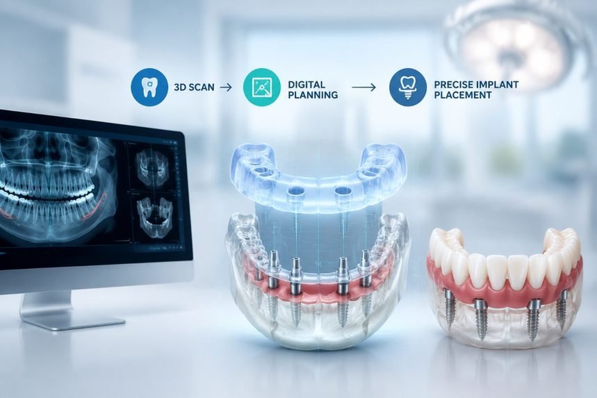
For most of its history, replacing a full mouth of teeth with implants meant committing to a plan that only fully revealed itself once surgery was already underway. Dentists worked from flat X-rays, a good deal of clinical instinct, and whatever they found after making the first incision. The results were usually solid, but the road there could be long and a little unpredictable.
Three-dimensional imaging changed the order of operations. The jaw is now mapped in full before any instrument touches it, so the surgery mostly confirms a plan rather than arriving at one. Someone typing full mouth dental implants near me into a search engine today is walking into a noticeably different experience than a patient who had the same work done a decade ago, even if the finished smile looks much the same.
The underlying biology has not moved. A titanium post is still placed in the jaw and left to fuse with the bone over several months, the gradual healing that clinicians call osseointegration. What has changed is nearly everything that happens before the drill starts.
From Educated Guesswork To A Digital Blueprint
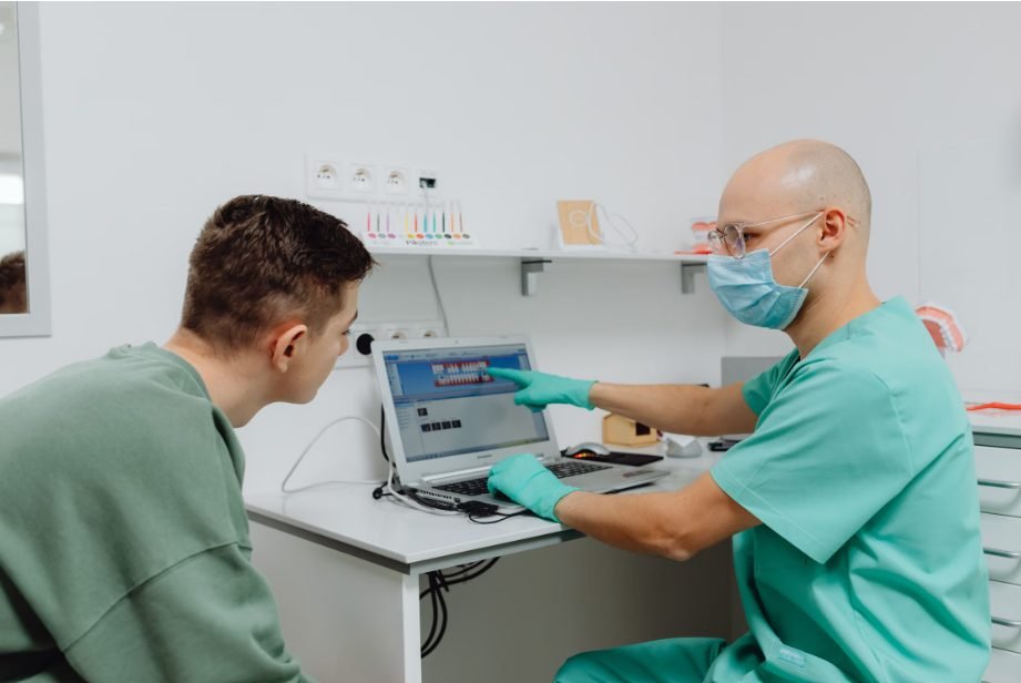
A cone beam CT scan gives the dental team a volumetric view of the jaw: bone height and width, the exact path of the nerves, the position of the sinuses, and how much solid bone is genuinely available at each planned implant site. From that scan, the whole case is rehearsed in software before the patient ever sits down for surgery. The plan can then be turned into a surgical guide, a custom template that fits over the remaining teeth or the gum and steers each drill to a set depth and angle.
The precision this buys is not hypothetical. Systematic reviews of the measurements taken from these scans have found they correspond closely to the real dimensions of the jaw, which is what makes them trustworthy enough to plan around. When those plans are carried out with a fully guided template, the implant tends to land within about a millimetre of where it was intended. A 2018 meta-analysis of static guided surgery put the average error near 1.2 mm at the entry point, and about 3.5 degrees of angulation, and a larger 2024 review pooling sixty-seven studies landed in the same territory, at roughly 1.1 mm at the crown, 1.4 mm at the tip, and again about 3.5 degrees off axis. Both point to the same conclusion: a fully guided protocol has become the benchmark for accuracy.
What Actually Changed, Side By Side
Sometimes the shift is easier to see laid out plainly than argued for.
| Stage | The Older Way | The 2026 Approach |
|---|---|---|
| Imaging | Flat, two-dimensional X-rays | A full 3D cone beam scan of the jaw |
| Planning | Worked out largely during surgery | Mapped digitally, well before the appointment |
| Placement | Positioned by hand and by eye | Driven by a printed or digital surgical guide |
| Healing | Longer and harder to predict | Often shorter and more consistent |
| Appointments | Usually more of them | Frequently fewer, since less is left to chance |
The distance between those two columns is not small. A dentist working from a 3D scan can read bone density, nerve position, and sinus location before ever picking up a handpiece. In the old workflow, much of that only became clear once the surgery had already begun.
Why So Much Rides On The Planning Stage
Full-mouth work is not a single procedure so much as a chain of them, and a full arch is unforgiving. Several implants have to sit in precise relation to one another and to the bridge of teeth they will eventually carry. Put one in the wrong place and the entire restoration can be thrown off. That is exactly the problem 3D planning was built to solve. The tricky spatial decisions get made and checked on a screen, where a mistake costs nothing, instead of in the chair, where it costs a great deal.
What The Shift Feels Like From The Patient’s Chair
For the person actually going through it, better imaging usually shows up as fewer surprises. Because the team already knows what it is working with, there tend to be fewer exploratory steps, tighter and more honest timelines, and appointments that account for healing and bone quality from the outset. Placement guided to a plan is often less invasive, which can mean less swelling and soreness afterward. Some practices can even show a patient a digital preview of the final result before anyone commits to treatment.
None of this makes every case straightforward. Bone loss, old dental work, smoking, and general health still shape how smoothly things go, and honest planning is partly about surfacing those complications early rather than pretending they are not there. What has genuinely improved is predictability.
Where The Money Goes, And Why
Full-mouth implants sit at the upper end of dental treatment costs, and 3D imaging is part of the reason, though not in a way that adds cost for its own sake. The scan and the digital plan are front-loaded expenses that tend to reduce the far more costly complications, revisions, and redo surgery that vague planning invites. A handful of factors move the final number more than the imaging does:
- How many implants are needed to support the arch
- Whether the bone needs grafting before implants can go in
- The material chosen for the final set of teeth
- The overall complexity of the case, including any existing dental work
Most clinics will only put a firm figure in writing after the scan and planning are done, because that is the first point at which the true scope of the work is clear. It also helps to remember what that money buys over time. Pooled data put the ten-year survival rate of contemporary implants at around 96 percent at the implant level, and reviews limited to studies with at least a decade of follow-up report similarly high survival and success. Those are the kind of long-term success rates that careful planning helps protect.
How To Tell Whether A Practice Has Truly Invested

Not every clinic has actually committed to this way of working, and the gap matters more than patients expect. A few plain questions tend to separate the practices that have from the ones that only talk a good game:
- Is a 3D cone beam scan a standard part of implant planning here, or something offered only now and then?
- Will the implants be placed with a surgical guide, or freehand?
- How many full-arch cases has the team actually completed?
- What is the plan if the scan shows less bone than expected?
Practices that have genuinely invested usually raise the imaging themselves, without being prompted. It is one reason patients comparing dental implants Virginia Beach practices increasingly ask about scanning and guided placement directly, rather than assuming every office offers the same thing.
Common Questions People Ask
How Long Does The Whole Process Take Now?
- It varies from person to person, but many full-mouth cases are completed within a few months rather than the longer, more drawn-out timelines older methods often required. The tighter planning that 3D imaging allows is a large part of why the schedule is more compressed and more predictable.
Is A 3D Scan More Expensive Than A Regular X-Ray?
- The scan itself usually costs more than a conventional X-ray. Across the full course of treatment, though, it often works out cheaper by lowering the odds of revision surgery and other expensive surprises.
Does 3D Imaging Make The Surgery Less Painful?
- Not directly, since the imaging does not numb anything; what it tends to do is allow more precise, less invasive placement, which is often associated with less tissue trauma and an easier recovery.
Can A Scan Tell Whether Someone Is A Candidate For Implants?
- To a large degree, yes. Bone density and structural issues show up clearly on a 3D scan, so the team can judge suitability and plan around any shortfall, sometimes by grafting first.
What About The Radiation From A Cone Beam Scan?
- Cone beam imaging does use ionizing radiation, so responsible clinics order a scan only when it will genuinely inform treatment and follow the principle of keeping the dose as low as reasonably achievable. Research also continues to push scanning protocols toward lower doses without losing the detail needed to plan.
How Do I Find A Dentist Who Actually Uses This?
- Ask directly whether a 3D cone beam scan is a standard part of planning rather than an add-on, and whether placement is guided. Offices that have truly adopted the technology tend to bring it up before you do.
Takeaway For Anyone Weighing Implants
3D imaging did more than make full-mouth implant treatment more accurate. It made the whole thing easier to see, for patient and dentist alike. Patients get a clearer picture of what lies ahead before committing, and clinicians work with a level of precision that flat X-rays could not offer. If you are considering this kind of treatment, the imaging setup is one of the more useful things to ask about in that first consultation, and it is well worth comparing a few practices specifically on how far each has gone with 3D planning and guided placement.
Medical Disclaimer
This article is for general information only and is not a substitute for professional dental or medical advice, diagnosis, or treatment. Individual circumstances vary, and no outcome can be guaranteed. Always consult a qualified dentist, oral surgeon, or other suitably licensed clinician about your own situation before making any treatment decision. Never disregard or delay seeking professional advice because of something you have read here.
References
- Fokas G, Vaughn VM, Scarfe WC, Bornstein MM. Accuracy of linear measurements on CBCT images related to presurgical implant treatment planning: a systematic review. Clinical Oral Implants Research. 2018;29(Suppl 16):393-415. https://doi.org/10.1111/clr.13142
- Tahmaseb A, Wu V, Wismeijer D, Coucke W, Evans C. The accuracy of static computer-aided implant surgery: a systematic review and meta-analysis. Clinical Oral Implants Research. 2018;29(Suppl 16):416-435. https://doi.org/10.1111/clr.13346
- Khaohoen A, Powcharoen W, Sornsuwan T, Chaijareenont P, Rungsiyakull C, Rungsiyakull P. Accuracy of implant placement with computer-aided static, dynamic, and robot-assisted surgery: a systematic review and meta-analysis of clinical trials. BMC Oral Health. 2024;24:359. https://doi.org/10.1186/s12903-024-04033-y
- Howe M-S, Keys W, Richards D. Long-term (10-year) dental implant survival: a systematic review and sensitivity meta-analysis. Journal of Dentistry. 2019;84:9-21. https://doi.org/10.1016/j.jdent.2019.03.008
- Yeung AWK, Jacobs R, Bornstein MM. Novel low-dose protocols using cone beam computed tomography in dental medicine: a review focusing on indications, limitations, and future possibilities. Clinical Oral Investigations. 2019;23(6):2573-2581. https://doi.org/10.1007/s00784-019-02907-y
- Moraschini V, Poubel LADC, Ferreira VF, Barboza EDSP. Evaluation of survival and success rates of dental implants reported in longitudinal studies with a follow-up period of at least 10 years: a systematic review. International Journal of Oral and Maxillofacial Surgery. 2015;44(3):377-388. https://doi.org/10.1016/j.ijom.2014.10.023